マウスの断面図、解剖のアトラスとして今回出版しました。すでに、「ウサギ編」「ラット編」が上梓されていることでもあり、先端医学の画像解析に必要となる「断面的・立体的な解剖学的知識」に益することを目的とした参考書です。本書は「マウスのマクロ解剖写真・水平断・矢状断及び接断の断面写真を『生体に近い状態の解剖アトラス』として、カラー図版(150面)により立体的に理解しやすいように工夫されたものです。
・マウス解剖図のカラー写真版としては世界で初めての本。
・図譜はカラー写真で構成し、細部まで検討できるよう拡大してある。
・無固定(凍結状態で切断、融解後撮影)で、より生体に近い状態の
カラーアトラスを作成。生体情報が手にとるようにわかる。
・解剖名にはすべて日本語と英語が併記してあり、辞書的な活用も
可能である。
著者
岩城隆昌 (東京慈恵会医科大学実験動物施設助教授) 山下 廣 (東京慈恵会医科大学第一解剖学教室教授) 早川 敏之(東京慈恵会医科大学第一解剖学教室助教授) 2012年5月2日 初版 第4刷発行 2015年11月 初版 第5刷発行
【特 色】
無固定で生体に近い状態の解剖カラーアトラス(断面解剖写真、マクロ解剖写真とも) 切断部分をわかりやすく例示 臓器名、組織名は日本語、英語の2か国語同時表記 断面写真はスケールで実寸を表示 細部まで鮮明なマクロ写真 細部まで十分に検討できるA4判カラー200ページ、掲載写真点数150点 検索しやすい日本語、英語の2か国語索引
【マウスの断面解剖アトラス はじめに】
マウスは古くから実験動物として使われ、20世紀初頭には研究目的に合わせて各種の近交系、ミュータント系、クローズドコロニーやそれらの交雑系が作られるまでに至っている。さらに近年、相同遺伝子組み換え技術を用いたヒト遺伝子の基礎的研究や解析、各種病気の解明・治療等の研究に遺伝子導入マウスや遺伝子破壊マウスが全世界で新たに産生されるようになり、今や近代医学をはじめとする生命科学研究に欠かせない動物となっている。遺伝子導入マウスや遺伝子破壊マウスは遺伝子の作用機構の解析上、個体の形態異常を正確に把握することは重要なステップと位置づけられ、そのため形態異常を的確に観察する能力や発見した異常が系統間の自然発生異常と異なると判断するための解剖学的な知識の要求が高まっている。一方、CT(computed axial tomography)、超音波エコー、MRI(magnetic resonance imaging)、SPECT(single photon computed tomography)やPET(positron emission tomography)等の画像診断装置の発達により、生体内の情報が無侵襲に手に入るようになり、小型の実験動物を用いた研究にも利用されるようになってきた。これらの画像解析には断面的・立体的な解剖学的知識が必要とされる関係から生体の情報が理解しやすいかたちの解剖学の参考書が要望されるようになってきている。 著者らは平成5年に、実験動物の断面解剖アトラス・ウサギ編(チクサン出版社・英語,ラテン語, 日本語併記)を、平成9年に実験動物の断面解剖アトラス・ラット編(チクサン出版社・英語,日本語併記)を出版した。今回これらの経験を生かし、上記要望に微力ながら応えることができれば幸いと考え、クローズドコロニー系(ICRやddY)マウスの解剖アトラスを出版することを決意した。本書はマウスのマクロ解剖写真と、水平断、矢状断および横断の断面写真を体系的に配したカラーアトラス集で、以下の点を留意して作成した。 1)無固定(断面画像は安楽死・脱毛処理後に-40℃近くで凍結した動物を、ダイヤモンドディスクソーを使用して切断、生理食塩液にて融解後に写真撮影)で生体に近い状態の解剖カラーアトラスを作成し、生体の断面情報を研究する研究者の参考に成り得ることを計った。 2)図は血管造影図(2ページ)を除き、全てカラー写真で構成(総図譜150ページ)し、しかも紙面の許す範囲で大型に配した。 3)断面解剖と平行して切断面のX線写真やマクロ写真を配して、体の中での位置的状態が立体的に理解しやすいように工夫した。 4)解剖名は日本語と英語を同時に表記し、読者が日本語と同時に英語の解剖名も把握できるように企画した。 本書の作成にあたり下記の著書を参考とした。 1) Cook, M.J. (1965).The Anatomy of the Laboratory Mouse. Academic Press, London. 2) Foster, H.L., J.D. Small and J.G. Fox(ed). (1983). The Mouse in Biomedical Research Vol.3 Chapter 7.Anatomy. Academic Press. London. 3) Green, E. L. (ed). (1966). Biology of the Laboratory Mouse. McGraw-Hill, New York. Chapter 13. Anatomy, Katharine P.H. et al. 4) Gude, W.D., G.E. Cosgrove and G.P. Hiesch. (1982). Histological Atlas of the Laboratory Mouse. Plenum Press. New York. 5) Kaufman, M.H. (1992). The Atlas of Mouse Development. Academic Press, London. 6) Popesko, P., V. Rajtova, and J. Horak. (1992). A Color Atlas of Anatomy of Small Laboratory Animals 2. Mouse. Wolfe Publishing Ltd., London. 7) Sidman, R.L., J.B. Angevine Jr. and E.T.Pierce. (1971). Atlas of the Mouse Brain and Spinal Cord. Harvard University Press, Cambridge. 8) Theiler, K. (1989). The House Mouse: Atlas of Embryonic Development. Springer-Verlag, New York. なお不適当な写真、用語等について読者諸賢のご批判を仰ぎ、今後の参考にさせて頂ければ幸いである。 謝辞 本書の出版にあたり以下の方々に多大なご指導ご援助を頂いた東京慈恵会医科大学医科学研究所実験動物施設 施設長 大川 清 教授、同施設 成相孝一 助手ならびに施設職員諸氏に感謝を申し上げる。また動物は我が国を代表する下記の実験動物生産・販売業者から分与または購入したものを自家繁殖して用いたが、その際、絶大なご援助・ご協力さらに資料の提供に応じて頂いた、株式会社 日本医科学動物資材研究所、日本クレア株式会社、日本チャールス・リバー 株式会社、日本エス エル シー株式会社および三共ラボ 株式会社の担当諸氏に心より感謝を申し上げると共に我が国の実験動物生産が今後とも益々発展することをお祈りする。 2001年 4月 著者ら記
本書を推薦する
慶應義塾大学医学部教授 前 島 一 淑 マウスは、いうまでもなく、生命科学研究に不可欠の実験素材であり、もっとも多く使われている実験動物である。 それだけに、マウスの形態学的なイメージをカラー写真と日・英語の解剖学名を付された本書の完成は高く評価されるべきである。 じつは、ご承知のこととは思うが、本書は慈恵医大の岩城隆昌博士のグループが実験動物の断面解剖アトラスを手がけ、上梓された「実験動物の断面解剖アトラス」(ウサギ編)、同じく「ラット編」に続く、第三弾目にあたる。また、このシリーズも今回で太尾となる。 既刊と同様、マウスの「矢状断、水平断、前頭断、その他の関連部位」についての「断面」映像を主としており、とくにマウスを用いる研究者、研究室や実験施設では、必備の図書であることはいうまでもない。 岩城博士等の努力によって貴重な「解剖アトラス・シリーズ」が完成したことを喜びたい。
目 次
目 次
Contents
はじめに
Introduction iii
骨格
Skeleton
全身骨格:左側観 1
Skeleton : left lateral view
骨格(頭部と前肢):左背側観 2
Skeleton (head and forefoot) : left dorsal view
頭蓋骨:背側観と腹側観 3
Skeleton of the head : dorsal and ventral surfaces
頭蓋骨:頭蓋冠(内面観)と内頭蓋底(内面観 ) 4
Skeleton of the head : view of the ventral endocranial skullcap and dorsal internal base of skull
骨格(四肢と椎骨):右側観 5
Skeleton (limb and vertebrae) : right lateral view
骨格(左上肢):左側観 6
Skeleton of the left thoracic limb : left lateral view
右後肢:足底観と背側観 7
Skeleton of the right pelvic limb : view of pantal and dorsal surfaces
寛骨:背側観と腹側観 8
Skeleton of coxae : view of the dorsal and ventral surfaces
頭部,顔面のマクロ写真と水平断
Head and Facies(Gross photographs and Dorsal sections)
頭部:背側観(頭蓋骨上から中大脳動脈, 横静脈洞, 静脈洞交会, 大脳半球等を観察) 9
Head : dorsal view of skull with skin removed
頭部:背側観(頭蓋骨および脳硬膜を除去して嗅脳,大脳半球,小脳等を観察) 10
Head : dorsal view of rhinencephalon, cerebral hemisphere and cerebellar hemisphere with bones of skull and entocranium removed
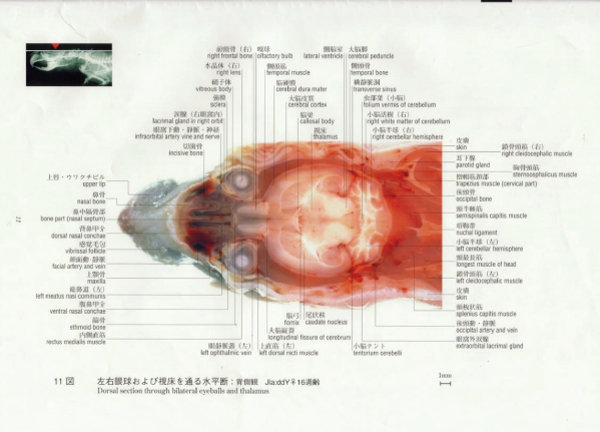
左右眼球および視床を通る水平断:背側観 11
Dorsal section through bilateral eyeballs and thalamus
鼻端と左右眼球を通る頭部水平断:背側観 12
Dorsal section of head through rostrum and bilateral eyeballs
ウワクチビルと左右眼球および視神経を通る頭部水平断:背側観 13
Dorsal section of head through upper lip and bilateral eyeballs, view through upper lip and bilateral optic nerves
硬口蓋と視神経および鼻腔と視床を通る頭部水平断:腹側観 14
Dorsal section through hard palate and optic nerve, through nasal cavity and thalamus : ventral views
眼球,視神経と下垂体:左側観(左側の前頭骨,頭頂骨,側頭骨,頬骨等を切除した) 15
Eyeball, optic nerve and pituitary : left lateral view
眼と眼静脈叢:左側観 16
Eye and ophthalmic vein : left lateral views
頭頚部,前肢のマクロ写真と矢状断
Head, neck and fore-limbs(Gross photographs and Sagittal section)
頭頚部および前肢:左右外側面(剥皮して観察) 17
Head, neck and fore-limbs : left and right lateral views (skin removed)
頭頚部:腹側観(剥皮して観察) 18
Head and neck : ventral view (skin removed)
頭頚部(剥皮して観察): 腹側観と左側観 19
Head and neck (skin removed) : ventral and left lateral views
頭頚部腹側観:気管と総頚動脈 20
Head and neck : ventral view of trachea and common carotid artery
甲状腺と上皮小体:腹側観と左側観 21
Thyroid gland and parathyroid gland : view of the ventral and left lateral surfaces
臼歯を通る右頭頚部矢状断:右側観と上下切歯 22
Sagittal section through right molar tooth : right lateral view and view of cranial and caudal incisor teeth
頸胸部,胸腔内と肺のマクロ写真
Neck, chest, thoracic cavity and lungs(Gross photographs)
胸部:腹側観 23
Neck and chest : view of ventral surface (skin removed)
胸腔内:左側観と右側観 24
Viscera of thoracic cavity from left side and right side : major part of left and right lungs removed
胸部腹側観(心臓と胸腺) 25
Viscera of thoracic cavity (heart and thymus) : ventral view
胸部右側観 26
Chest : view of right lateral surface and thoracic cavity from right side

肺の背側観:胸腔を開けて観察(右図は肺胞内に空気を入れた) 27
Lungs in thoracic cavity : dorsal view (air pumped into trachea in right photograph)
肺:背側観と腹側観(左右共に肺胞内に空気を入れた) 28
Lungs and heart : view of the dorsal and ventral surfaces (air pumped into trachea)
胸部と腹部臓器のマクロ写真
Thoracic and abdominal organs(Gross photographs)
胸部と腹部の内臓(心臓,肝臓,胃,腎臓と胸大動脈):腹側観 29
Male thoracic and abdominal organs (heart, liver, stomach, kidney and thoracic aorta) : view of ventral surface
腹部消化管(下行結腸,直腸,肛門):腹側観 30
Abdominal organs (descending colon, rectum and anus) : view of the ventral surface
腹腔の臓器:肝臓,消化管と膀胱(排尿後) 31
Abdominal viscera : liver, intestinal tracts and urinary bladder (after urination)
腹部臓器:左側観 と右側観(肝臓,胃,脾臓,卵巣) 32
Female abdominal cavities (liver, stomach, spleen and ovary) : view from the left side and from the right side
肝臓:横隔面と臓側面 33
Liver : diaphragmatic surface and visceral surface
肝臓:胆嚢, 総胆管と十二指腸:腹側観 34
Liver : gall bladder, common bile duct and duodenum : ventral view
門脈:腹側観 35
Portal vein : view of the ventral surface
肝臓:横隔面と臓側面(♀13週齢より摘出) 36
Liver : view of diaphragmatic and visceral surfaces
腹腔の臓器(腸間膜リンパ節,空腸動静脈,回腸動静脈,前腸間膜静脈) 37
Abdominal viscera : mesenteric lymph node, jejunal artery and vein, ileal artery and vein
消化管(舌から肛門まで) 38
Digestive tract (removed and displayed)
胃と十二指腸,胃(胃粘膜面)および 幽門から十二指腸にかけての拡大 39
Stomach and duodenum : view of cranial surface, longitudinal sections of mucous tunica of stomach and pyloric part of stomach
腎臓と副腎 40
Kidney and suprarenal glands : dextral, sinistral and bilateral
尿管と膀胱: 尿管を現すため後方に持ち上げた膀胱と脂肪に囲まれた雌の膀胱 41
Ureter and urinary bladder : bladder was turned up to show running ureters
雌雄生殖器のマクロ写真
Male and female reproductive organs(Gross photographs)
雄外性器:腹側観(包皮を下方に反転,陰嚢を除去) 42
Ventral view of male external genitalia (prepuce turned down, scrotal sacs removed)
雄生殖器(摘出):腹側観 43
Male reproductive organs (removed and displayed) : ventral view
腎臓と卵巣:腹側観と背側観 44
Kidneys and ovaries : ventral view and dorsal view
雌性殖器:開腹(腎上部へ左卵巣を移動して観察,摘出臓器):腹側観 45
Female genital organs : abdominal cavity, ovary and uterine tube, enucleated, ventral views
腟,膀胱,尿管と外陰部(妊娠末期) 46
Vagina, bladder, urethra of female and feminine pudendum (gravid uterus)
子宮,子宮頸部と腟(経産) 47
Multiparous of uterus, cervix and vagina in situ (transverse section through vagina to interior part of uterus)
妊娠母獣と胎仔のマクロおよび断面写真
Pregnant females and fetuses(Gross and sectional photographs)
妊娠母獣腹側観(交尾後20日) 48
Ventral view of pregnant female 20 days after mating
妊娠母獣右側矢状断(交尾後20日) 49
Sagittal section of pregnant female 20 days after mating : right lateral view
妊娠母獣水平断(妊娠末期):腹側観 50
Dorsal section through pregnant uterus (late pregnancy)
妊娠母獣前頭断(妊娠末期):尾側観 51
Transverse section through the pregnant uterus (late pregnancy) : caudal views
妊娠母獣横断面(妊娠末期):尾側観 52
Transverse sections through pregnant uteri (late pregnancy) : caudal views
妊娠子宮(妊娠末期) 53
Pregnant female with uterus displayed (late pregnancy)
妊娠子宮と胎仔(雄と交尾後20日経過):腹側観 54
Pregnant uteri and fetuses : vagina and body of uteri were reversed upward, parts of uterus and fetus amnios have been cut away
胎仔と胎盤 (雄と交尾後20日経過) 55
Fetuses and placenta : vagina and the body of uterus were reversed upward after extirpation and macrograph of fetuses
体表(背側観と腹側観)のマクロ写真
Dorsal and ventral view of superficial bodies(Gross photographs)
体表の浅層の筋:背側観 56
Superficial muscles of the body : dorsal view
体表の浅層の筋:腹側観 57
Superficial muscles of the body : ventral view
四肢と尾のマクロ写真
Limbs and tail(Gross photographs)
後肢外表面:左側観と 腹側観 58
Limbs : superficial layer, view of left lateral and ventral
後肢(腹側観と左側観)と尾の表在動静脈(腹側観と背側観) 59
Artery and vein of pelvic limb and tail : ventral, left lateral and dorsal views
四肢:前肢 (背側観と尾側観)および後肢(内側観と尾側観) 60
The limbs : dorsal view and caudal view of forearms, medial view and caudal view of hindpaws
乳腺のマクロ写真
Mammary glands(Gross photographs)
乳腺:背側観 61
Mammary gland : dorsal view
乳腺:左側および右側観 62
Mammary gland : left and right lateral views
乳腺:腹側観 63
Mammary gland : ventral view
新生仔の外尿道口
external urethral openings(new born mice)
雌雄新生仔の外尿道口:尾側観 64
Male and female external urethral openings (newborn mice)
矢状断(右側観と左側観)
Sagittal sections(right and left lateral views)
右上腕と右大腿表面を通る矢状断:右側観 65
Sagittal section through right humerus and anterior surface of right femur : right lateral view
右咬筋と右大腿骨を通る矢状断:右側観 66
Sagittal section through right masseter muscle and right distal end of femur : right lateral view
右咬筋浅部と右大腿四頭筋を通る矢状断:右側観 67
Sagittal section through lower part of right masseter muscle and right quadriceps femoris muscle : right lateral view
右頬骨弓と右膝関節を通る矢状断:右側観 68
Sagittal section through right zygomatic arch and right knee joint : right lateral view
右眼球と右尾根部を通る矢状断:右側観 69
Sagittal section through right eyeball and right lateral root of tail : right lateral view
右眼球と右膝関節を通る矢状断:右側観 70
Sagittal section through right eyeball and right knee joint : right lateral view
右肩関節と右大腿骨を通る矢状断:右側観 71
Sagittal section through right shoulder joint and right femur : right lateral view
右下唇と右膝蓋骨を通る矢状断:右側観 72
Sagittal section through right lower lip and right patella : right lateral view
右腹鼻甲介と右副腎を通る矢状断:右側観 73
Sagittal section through right ventral nasal conchae and right suprarenal gland : right lateral view
鼻骨から直腸を通る正中矢状断:右側観 74
Midsagittal section through right nasal bone and rectum : right lateral view
正中矢状断鏡面像(頭部から胸部):右側観と左側観 75
Midsagittal section through rostrum of nose and liver : mirror symmetry views, right and left lateral views
左眼球と副腎を通る矢状断:右側観 76
Sagittal section through left eyeball and suprarenal gland : right lateral view
上唇から肛門を通る正中矢状断:左側観 77
Midsagittal section through right upper lip and anus : left lateral view
右外鼻孔と右腎臓を通る矢状断:左側観 78
Sagittal section through right external naris and right kidney : left lateral view
左鼻中隔と直腸を通る矢状断:左側観 79
Sagittal section through left nasal septum and rectum : left lateral view
左眼球(硝子体)と左腎臓を通る矢状断:左側観 80
Sagittal section through the left vitreous body and left kidney : left lateral view
下垂体,胸腺と左腸骨翼を通る矢状断:左側観 81
Sagittal section through pituitary, thymus and left ala ossis ilii : left lateral view
左咬筋と左大腿骨頭を通る矢状断:左側観 82
Sagittal section through masseter muscle and head of left femur : left lateral view
左外耳道, 左腎臓および左大腿二頭筋を通る矢状断:左側観 83
Sagittal section through left external auditory meatus, left kidney and left biceps femoris muscle : left lateral view
左肘関節と左大腿骨を通る矢状断:左側観 84
Sagittal section through left elbow joint and left femur : left lateral view
水平断(背側観と腹側観)
Dorsal sections(dorsal and ventral views)
僧帽筋頚部と仙骨棘突起を通る水平断:背側観 85
Dorsal section through trapezius muscle of cervical part and spinous process of sacrum : dorsal view
脳梁幹と椎体を通る水平断:背側観 86
Dorsal section through trunk of corpus callosum and bodies of vertebra : dorsal view
第三脳室,肛門と精巣を通る水平断:背側観 87
Dorsal section through third ventricle, anus and bilateral testes : dorsal view
左右眼球および下垂体を通る水平断:背側観 88
Dorsal section through bilateral eyeballs and pituitary : dorsal view
鼻吻と 肛門 を通る水平断:背側観 89
Dorsal section through rostrum of nose and anus : dorsal view
鼻尖,左右副腎と腎臓を通る水平断:背側観 90
Dorsal section through nasal apex, bilateral suprarenal glands and bilateral kidneys : dorsal view
舌体,後大静脈と左右腎臓を通る水平断:背側観 91
Dorsal section through body of tongue caudal vena cava and bilateral kidney : dorsal view
舌根と直腸を通る水平断:背側観 92
Dorsal section through roof of tongue and rectum : dorsal view
喉頭蓋,後大静脈と肛門を通る水平断:腹側観 93
Dorsal section through epiglottis, caudal vena cava and anus : ventral view
鼻中隔軟骨部と左右卵巣を通る水平断:腹側観 94
Dorsal section through cartilage part of nasal septum and bilateral ovaries : ventral view
外頚静脈と大腿部を通る水平断:腹側観 95
Dorsal section through external jugular vein and femoral region : ventral view
舌体と外陰部(雌の)を通る水平断:腹側観 96
Dorsal section through body of tongue and feminine pudendum : ventral view
下顎骨と外陰部(雌の)を通る水平断:腹側観 97
Dorsal section through mandible and feminine pudendum : ventral view
精巣上体と精巣:水平断(背側観 )と開腹・剥皮像(腹側観) 98
Epididymides and testes : dorsal view(dorsal section)and ventral view(scrotal sacs removed)
横断面(吻側観と尾側観)
Transverse sections(rostral and caudal views)
鼻先の横断面:吻側観 99
Transverse sections through tips of noses : rostral views
左右の眼球を通る横断面:吻側観 100
Transverse section through bilateral eyeballs : rostral view
左右の水晶体を通る横断面:吻側観 101
Transverse section through bilateral lenses : rostral view
嗅球と眼窩内涙腺を通る横断面:尾側観 102
Transverse section through bilateral olfactory bulbs and intraorbital lacrimal glands : caudal view
嗅球と下切歯を通る横断面:尾側観 103
Transverse section through bilateral olfactory bulbs and caudal incisor tooth : caudal view
眼球後方を通る横断面:吻側観 104
Transverse section through post orbital part : rostral view
左右の視床下部皮質路と舌根部を通る横断面:吻側観 105
Transverse section through bilateral hippocampocortical tracts and root of tongue : rostral view
左右の下切歯根と眼窩内涙腺を通る横断面:尾側観 106
Transverse section through bilateral caudal incisor teeth and intraorbital lacrimal glands : caudal view
顎関節を通る横断面:尾側観 107
Transverse section through bilateral temporomandibular joints : caudal view
視床下部,下垂体を通る頭部横断面:尾側観 108
Transverse section through hypothalamus and pituitary : caudal view
視床,視床下部と舌骨体を通る断面図:尾側観 109
Transverse section through thalamus, hypothalamus and body of hyoid bone : caudal view
左右鼓室胞と咽頭を通る横断面:尾側観 110
Transverse section through bilateral tempanic bullae and pharynx : caudal view
左右の咽頭円蓋と顎下腺を通る横断面:吻側観 111
Transverse section through bilateral pharyngeal fornix and submandibular gland : rostral view
左右の内側皮質視床下部路と顎関節を通る横断面:尾側観 112
Transverse section through bilateral medial corticohypothalamic tract and temporomandibular joint : caudal view
左右梨状葉 と口峡を通る横断面:尾側観 113
Transverse section through bilateral piriform lobes and fauces : caudal view
松果体と蝸牛を通る横断面:吻側観 114
Transverse section through pineal body and cochlea : rostral view
左右の小脳半球と咽頭円蓋を通る横断面:吻側観 115
Transverse section through bilateral cerebellar hemispheres and pharyngeal fornices : rostral view
環椎と顎下腺を通る横断面:尾側観 116
Transverse section through atlas and bilateral submandibular glands : caudal view
第四頚椎と左右の上腕骨体を通る横断面:吻側観 117
Transverse section through 4th cervical vertebra and bilateral humeri : rostral view
第二胸椎と胸骨柄を通る横断面:尾側観 118
Transverse section through 2nd thoracic vertebra and manubrium of sternum : caudal view
第五胸椎と第四胸骨片を通る横断面:尾側観 119
Transverse section through 5th thoracic vertebra and 4th sternebra : caudal view
第六胸椎と第二胸骨を通る横断面:尾側観 120
Transverse section through 6th thoracic vertebra and 2nd sternum : caudal view
第七胸椎棘突起と第五胸骨片を通る横断面:尾側観 121
Transverse section through spinous process of 7th thoracic vertebra and 5th sternebra : caudal view
第八肋骨と左心室を通る横断面:尾側観 122
Transverse section through 8th right rib and left ventricle : caudal view
第十胸椎と心尖,剣状胸骨を通る横断面:尾側観 123
Transverse section through 10th thoracic vertebra, apex of heart and xiphisternum : caudal view
第十/十一胸椎椎間円板と胆嚢底を通る横断面:尾側観 124
Transverse section through 10th to 11th intervertebral disk of thoracic vertebrae and fundus of gall bladder : caudal view
第十一胸椎と剣状軟骨を通る横断面:尾側観 125
Transverse section through 11th thoracic vertebra and xiphoid cartilage : caudal view
第十一胸椎棘突起,胆嚢と剣状軟骨を通る横断面:尾側観 126
Transverse section through spinous process of 11th thoracic vertebra, gall bladder and xiphoid cartilage : caudal view
第十一胸椎棘突起と左右の第七肋軟骨を通る横断面:尾側観 127
Transverse section through spinous process of 11th thoracic vertebra and bilateral 7th costal cartilage : caudal view
第二腰椎と左副腎を通る横断面:尾側観 128
Transverse section through 2nd lumbar vertebra and left suprarenal gland : caudal view
第二腰椎,脾臓と右腎臓を通る横断面:尾側観 129
Transverse section through 2nd lumbar vertebra, spleen and right kidney : caudal view
第二/三腰椎椎間円板と左右腎洞を通る横断面:尾側観 130
Transverse section through 2nd to 3rd intervertebral disk of lumbar vertebra and bilateral renal sinus (kidney) : caudal view
第四腰椎と左右腎臓を通る横断面:尾側観 131
Transverse section through 4th lumbar vertebra and bilateral kidneys : caudal view
第四腰椎椎体と左右の腎臓を通る横断面:尾側観 132
Transverse section through vertebral body of 4th lumbar vertebra and bilateral kidneys : caudal view
第四腰椎後関節突起と左右腎臓を通る横断面:尾側観 133
Transverse section through caudal articular process of 4th lumbar vertebra and bilateral kidneys : caudalview
第四腰椎椎体と左右の卵巣静脈を通る横断面:尾側観 134
Transverse section through vertebral body of 4th lumbar vertebra and bilateral ovarian veins : caudal view
第六腰椎椎体と左右の精嚢腺を通る横断面:尾側観 135
Transverse section through vertebral body of 6th lumbar vertebra and bilateral vesicular glands : caudal view
第二仙椎,左右の精嚢腺と膀胱を通る横断面:尾側観 136
Transverse section through 2nd sacral vertebra, bilateral vesicular gland and urinary bladder : caudal view
第四仙椎と尿道(骨盤部:雄)を通る横断面:尾側観 137
Transverse section through 4th sacral vertebra and pelvic part of urethra of male : caudal view
第一尾椎と尿道海綿体を通る横断面:尾側観 138
Transverse section through 1st caudal vertebra and cavernous body of penis : caudal view
第一尾椎 椎孔と凝固腺を通る横断面:尾側観 139
Transverse section through vertebral foramen of 1st caudal vertebra and coagulational gland : caudal view
第一尾椎椎孔と包皮腺を通る横断面:尾側観 140
Transverse section through vertebral foramen of 1st caudal vertebra and bilateral preputial gland : caudal view
第一尾椎と恥骨結合を通る横断面:尾側観 141
Transverse section through 1st caudal vertebra and pubic symphysis : caudal view
第二尾椎と陰茎骨を通る横断面:尾側観 142
Transverse section through 2nd caudal vertebra and penis bone : caudal view
第二,第三尾椎間を通る横断面:尾側観 143
Transverse section between 2nd and 3rd caudal vertebrae : caudal view
第三尾椎と陰茎骨を通る横断面:尾側観 144
Transverse section through 3rd caudal vertebra and penis bone : caudal view
第三尾椎と包皮腺を通る横断面:尾側観 145
Transverse section through 3rd caudal vertebra and preputial gland : caudal view
第四尾椎椎体と肛門を通る横断面:尾側観 146
Transverse section through vertebral body of 4th caudal vertebra and anus : caudal view
第四尾椎と脛骨体を通る横断面:尾側観 147
Transverse section through 4th caudal vertebra and bilateral tibiae : caudal view
第六尾椎椎体と第四尾椎椎体を通る横断面:尾側観 148
Transverse sections through 6th and 4th vertebral body of caudal vertebrae : caudal views
血管造影 図(左側観と腹側観)
Angiogram(left lateral and ventral views)
血管造影 図:左側観 149
Angiogram : left lateral view
血管造影図:腹側観 150
Angiogram : ventral view
付録 Appendix 151
索引(日本語) Index(Japanese) 169
索引(英語) Index(English) 177